By Dr. Matthew G. Field, MD, PhD, MPH | Twin Cities Eye Consultants
When your vision becomes blurry, the natural response is to assume you need a new glasses prescription. But when prescriptions keep changing—or glasses never seem to bring things into focus—it is worth looking more closely at the cornea.
As a corneal specialist, I approach keratoconus with two goals: preserve the health and stability of the cornea and help each patient achieve the clearest vision possible. Recognizing the condition early gives us more opportunities to accomplish both.
Even with keratoconus, individuals can achieve remarkable success in careers and activities that place high demands on vision. A notable example is NBA superstar Stephen Curry, who has openly shared his experience with keratoconus. He has helped raise awareness of the condition through public advocacy efforts aimed at promoting earlier diagnosis and treatment, which can improve long-term outcomes for affected individuals.

What Is Keratoconus?
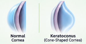
The cornea is the clear front window of the eye. Its smooth, rounded shape helps focus light onto the retina. In keratoconus, the cornea becomes thinner and bulges outward, developing an irregular, cone-like shape.
That irregularity disrupts how light enters the eye. Patients may experience blurred vision, ghost images, glare, or increasing astigmatism. Keratoconus usually affects both eyes, although one eye can be substantially worse than the other. It commonly becomes apparent during adolescence or young adulthood.
Signs That Deserve a Closer Look
Any of the following symptoms can be a sign you have keratoconus: frequent prescription changes, blurred or distorted vision, ghosting or double images, difficulty seeing at night, and vision that remains unsatisfactory despite glasses or contact lenses. But, remember, things can be very subtle at the beginning. So, if you are a teenager or young adult and aren’t correcting to 20/20 or have significant astigmatism particularly worse in one eye, I recommend being evaluated for this condition. A family history of keratoconus and frequent eye rubbing also deserve attention.
These clues do not automatically mean you have keratoconus. However, when they occur together, I recommend checking the cornea rather than simply updating the prescription again.
Why Does Keratoconus Develop?
The exact cause of keratoconus remains unclear and is an active area of research. A family history can increase risk, and the condition is also associated with Down syndrome and allergic eye disease.
However, most patients I see have no family history or identifiable genetic cause. I have also noticed an association with sleep apnea, so I routinely ask about sleep quality and recommend sleep testing when indicated. Repeated eye rubbing is an important modifiable risk factor, as is avoiding pressure on the eyes during sleep.
Early detection and timely treatment are essential to slowing progression, preserving the cornea, and protecting vision.
Why Early Diagnosis Matters
Keratoconus can progress before patients realize how much their vision has changed, especially when one eye compensates for the other. Because we cannot reverse the condition once it has progressed, the best opportunity to preserve vision is before significant corneal distortion or scarring occurs. Rate and degree of progression varies among individuals, so follow-up and treatment decisions should be tailored to each patient. In advanced cases with severe scarring or inadequate functional vision, a partial- or full-thickness corneal transplant may be required. My goal is to prevent patients from ever reaching that stage.
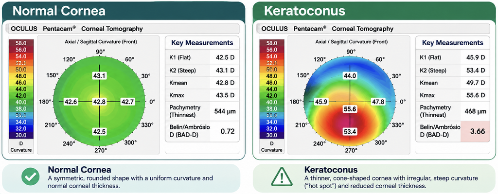
Evaluation includes a comprehensive eye examination and specialized imaging. Corneal tomography is essential for detecting and diagnosing keratoconus, providing detailed information about corneal shape and thickness. Clinically validated software algorithms can identify eyes that may be at risk, and comparing scans over time helps determine whether the disease is progressing.
How Do We Stop Progression?
One of the most useful distinctions in keratoconus care is the difference between improving vision and preventing further disease progression.
Glasses and soft contact lenses may help improve vision in early disease. As the cornea becomes more irregular, rigid gas-permeable, hybrid, or scleral lenses may provide better improvement in vision. However, these treatment modalities DO NOT stop progression of disease. Corneal specialists aspire to detect keratoconus at the earliest stages, so we can treat you before you even need specialized lenses.
The only treatment that currently exists to stop progression of keratoconus is collagen cross-linking.
Epithelium-On Cross-linking to Stop Disease Progression
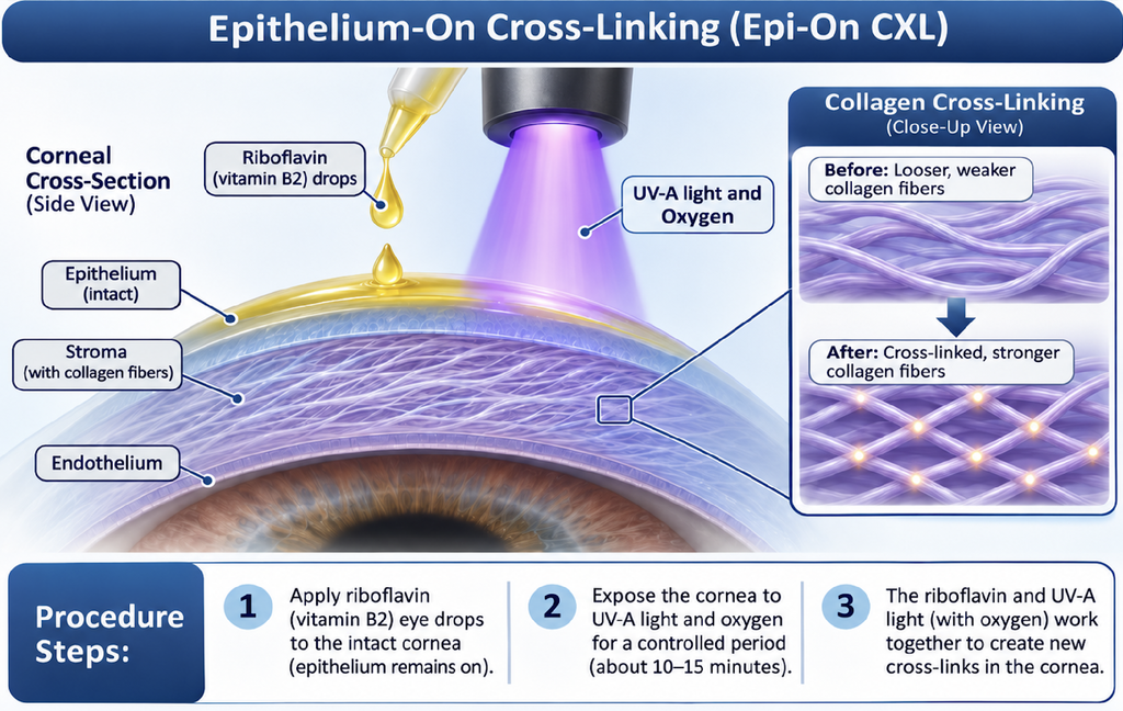
Keratoconus progresses when the collagen fibers that give the cornea its strength weaken, causing the cornea to thin and bulge forward. Corneal collagen cross-linking uses riboflavin (vitamin B2), ultraviolet A light, and oxygen to create additional bonds within the cornea, strengthening its structure. The primary goal is to halt or slow progression and preserve vision. While improved vision is not guaranteed, many patients experience modest visual improvement due to corneal flattening after treatment.
In October 2025, the FDA approved Epioxa, the first FDA-approved epi-on treatment of keratoconus. We have enrolled many patients in the traditional epi-off collagen cross-linking procedure with excellent results and are now excited to offer epi-on cross-linking to our patients! We will be enrolling our first patients with this new treatment modality this month, November 2026!
“Epithelial-on,” or “epi-on,” means the cornea’s outer epithelial layer remains in place. Traditional epithelial-off treatment removes that layer to facilitate riboflavin penetration. But epi-on treatment is faster, is less painful and has a quicker recovery with similar long-term efficacy results as epi-off cross-linking.
Keeping the surface intact does not eliminate recovery or risk. Redness, discomfort, light sensitivity, haze, epithelial defects, and reduced vision can occur.
When discussing cross-linking, I emphasize realistic expectations: its main value is protecting future vision, and glasses or contact lenses may still be necessary afterwards to get the best vision possible.
Corneal Tissue Addition Keratoplasty (CTAK) to Improve Vision and Corneal Shape
Many patients ask me if there is anything that they can do to improve their vision with keratoconus. They want to reduce their need for thick high-prescription glasses and/or specialized contact lenses. Excitingly, in the last couple of years corneal tissue addition keratoplasty (CTAK) has been developed.
CTAK addresses the irregular shape that makes vision difficult to correct. The surgeon uses a femtosecond laser to create a channel within the patient’s cornea. A customized piece of donor corneal tissue that is designed specifically for the patient is inserted into that channel that acts like a belt or girdle to help flatten and regularize the cornea while preserving the patient’s central tissue.
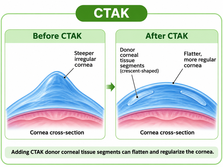
Results from clinical studies and my own experience have been very encouraging. While CTAK is not expected to restore perfect 20/20 vision, it can improve corneal shape and enhance visual quality, both with and without glasses or contact lenses.
I consider CTAK for selected patients whose vision remains limited by corneal irregularity. It may also be helpful before cataract surgery, as improving corneal shape can expand lens options and optimize post-cataract surgery visual outcomes.
CTAK can improve vision, but patients may still need glasses or specialty contact lenses. It also does not replace corneal cross-linking when progression is present. Cross-linking improves corneal stability and halts disease progression, while CTAK improves corneal shape. Because these treatments complement each other, I often perform them within a few weeks of one another.
If you have signs or symptoms of keratoconus, talk with your eye doctor or contact our office. Early detection and treatment can prevent progression and preserve vision, and newer therapeutic options, such as CTAK, can help improve vision in patients with more advanced disease. My goal is to help every patient achieve the best vision possible.